43 simple microscope diagram with labels
Microscope labeled diagram - SlideShare Microscope labeled diagram 1. The Microscope Image courtesy of: Microscopehelp.com Basic rules to using the microscope 1. You should always carry a microscope with two hands, one on the arm and the other under the base. 2. You should always start on the lowest power objective lens and should always leave the microscope on the low power lens ... Label the microscope — Science Learning Hub All microscopes share features in common. In this interactive, you can label the different parts of a microscope. Use this with the Microscope parts activity to help students identify and label the main parts of a microscope and then describe their functions. Drag and drop the text labels onto the microscope diagram.
Simple Microscope - Definition, Diagram, FAQs Define Microscope. Simple Microscope Definition: A Simple Microscope meaning is used to see a magnified image of an object. Antonie Van Leeuwenhoek, a Dutchman, invented the first simple microscope, consisting of a single powerful magnetic lens that rotates to detect tiny freshwater insects. It is composed mainly of light microscopes.
Simple microscope diagram with labels
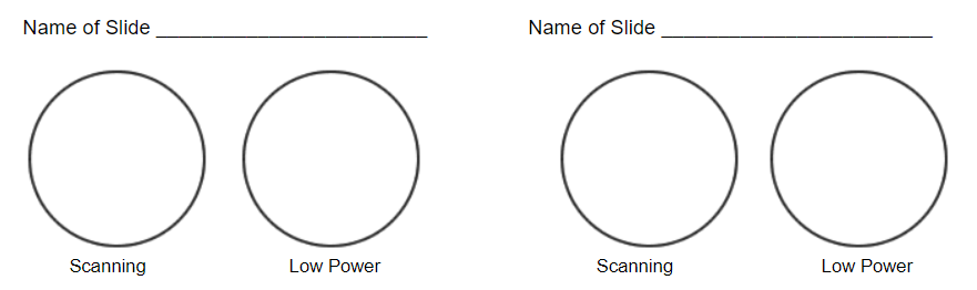
Simple Microscope - Diagram (Parts labelled), Principle, Formula and Uses Parts of a Simple Microscope A simple microscope consists of Optical parts Mechanical parts Labeled Diagram of simple microscope parts Optical parts The optical parts of a simple microscope include Lens Mirror Eyepiece Lens A simple microscope uses biconvex lens to magnify the image of a specimen under focus. MasteringBiology: Ch 10 Flashcards & Practice Test | Quizlet Drag the labels from the left to their correct locations in the concept map on the right. Not all labels will be used. ... Chloroplast membrane vesicles are equilibrated in a simple solution of pH 5 . The solution is then adjusted to pH 8 . ... Drag the labels to the appropriate locations on the diagram of the thylakoid membrane. red top ... Microscope Labeling - The Biology Corner 1) Start with scanning (the shortest objective) and only use the COARSE knob . Once it is focused… 2) Switch to low power (medium) and only use the COARSE knob . You may need to recenter your slide. Once it is focused.. 3) Switch to high power (long objective).
Simple microscope diagram with labels. Types of Microscopes: Definition, Working Principle, Diagram ... Where, D is the least distinct vision; F is the focal length of the convex lens; Simple Microscope Diagram. Principle of Simple Microscope. The working principle of a simple microscope is that when a sample is placed within the focus of the microscope, a virtual, erect and magnified image is obtained at the least distance of distinct vision from the eye that is held at the lens. › books › NBK21116Mapping Genomes - Genomes - NCBI Bookshelf Draw a diagram showing how a double-stranded cDNA is synthesized. 15. Define the term ‘mapping reagent’ and explain how a panel of radiation hybrids is used as a mapping reagent. 16. Explain how a clone library is used as a mapping reagent. 17. Draw a diagram to show how a sample of a single human chromosome can be obtained by flow cytometry. Parts of the Microscope with Labeling (also Free Printouts) Parts of the Microscope with Labeling (also Free Printouts) A microscope is one of the invaluable tools in the laboratory setting. It is used to observe things that cannot be seen by the naked eye. Table of Contents 1. Eyepiece 2. Body tube/Head 3. Turret/Nose piece 4. Objective lenses 5. Knobs (fine and coarse) 6. Stage and stage clips 7. Aperture pages.zeiss.com › rs › 896-XMS-794Principles of Fluorescence and Fluorescence Microscopy - ZEISS The Fluorescence Microscope The main requirement of a fluorescence microscope is to illuminate a specimen with light of an excitatory wavelength whilst simultaneously collecting and separating the compara-tively weaker light emitted by the sample. In the example of Stokes’ observation, these tasks are performed by the
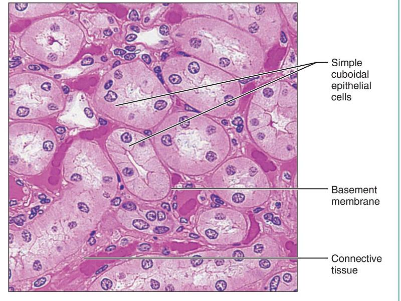
Anatomy Chart - How to Make Medical Drawings and Illustrations Pathologic anatomy focuses on how diseases affect and change the human body. Histology studies microscopic anatomy such as tissues and cells visible only under a microscope. Anatomy charts serve two main purposes: education in the form of anatomy worksheets and presentation in the form of simple healthcare illustrations. Simple Microscope - Definition, Types, Working Principle & Formula A simple microscope consists of a convex lens of a short focal length. The below figure shows the ray diagram which subsequently forms the image of an object (or we can say a source of light). (Image will be Updated soon) F is the focal length of the lens. An object is placed between the focal length and the centre of the curvature. Microscope Poster - Diagram with Labels | Teach Starter A poster containing a diagram with labels showing the key parts of a microscope. In Science it is important that students know how to use a variety of tools when conducting scientific experiments and inquiry. This poster focuses on the microscope and highlights its key parts. There are two print options available for this poster: Parts of a Microscope Labeling Activity - Storyboard That Create a poster that labels the parts of a microscope and includes descriptions of what each part does. Click "Start Assignment". Use a landscape poster layout (large or small). Search for a diagram of a microscope. Using arrows and textables label each part of the microscope and describe its function. Copy This Storyboard* More options
Compound Microscope Parts, Functions, and Labeled Diagram Compound Microscope Definitions for Labels. Eyepiece (ocular lens) with or without Pointer: The part that is looked through at the top of the compound microscope. Eyepieces typically have a magnification between 5x & 30x. Monocular or Binocular Head: Structural support that holds & connects the eyepieces to the objective lenses. Parts of a microscope with functions and labeled diagram Figure: Diagram of parts of a microscope There are three structural parts of the microscope i.e. head, base, and arm. Head - This is also known as the body. It carries the optical parts in the upper part of the microscope. Base - It acts as microscopes support. It also carries microscopic illuminators. Parts of a Simple Microscope - Labeled (with diagrams) Parts of a Simple Microscope - Labeled (with diagrams) A simple microscope is a very first type of microscope ever created. It consists of simple parts and performs simple functions. Although there are now many advanced microscope types, some applications may still demand the use of a simple microscope. Fluorescence Resonance Energy Transfer (FRET) Microscopy Presented in Figure 3 is a Jablonski diagram illustrating the coupled transitions involved between the donor emission and acceptor absorbance in fluorescence resonance energy transfer. Absorption and emission transitions are represented by straight vertical arrows (green and red, respectively), while vibrational relaxation is indicated by wavy ...
Microscope Introduction – “e” Lab - Biology LibreTexts
quizlet.com › 568033559 › botany-exam-1-chs-1/2/3-4Botany Exam 1 Chs. 1, 2, 3, 4, 5, 6, 7, 12, and 16 Quizzes Mitosis, or the division of a mother cell's nucleus into two identical daughter nuclei, is typically divided into four phases. Match each of the labels to identify what events take place during each phase of mitosis. 1. Prophase 2. Metaphase 3. Anaphase 4. Telophase A) Chromosomes are aligned at the equator of the cell and the spindle is fully ...
Print Anatomy Midterm 2 flashcards | Easy Notecards
› 2022 › 03Inside the Apple-1's unusual MOS clock driver chip Mar 21, 2022 · The six-bit display characters were held in six 1024-bit shift registers. A seventh shift register tracked the cursor position. 3 The diagram below shows the shift registers and the clock driver on the Apple-1 circuit board. These chips are in 8-pin packages, so two chips fit into the space of a regular TTL chip.

HistoLab2.htm
Labelled Diagram of Compound Microscope - Biology Discussion The below mentioned article provides a labelled diagram of compound microscope. Part # 1. The Stand: The stand is made up of a heavy foot which carries a curved inclinable limb or arm bearing the body tube. The foot is generally horse shoe-shaped structure (Fig. 2) which rests on table top or any other surface on which the microscope in kept.
Biology Microscope Diagram Gcse - Micropedia
Compound Microscope- Definition, Labeled Diagram, Principle, Parts, Uses The optical microscope often referred to as the light microscope, is a type of microscope that uses visible light and a system of lenses to magnify images of small subjects. There are two basic types of optical microscopes: Simple microscopes. Compound microscopes. The term "compound" in compound microscopes refers to the microscope having ...
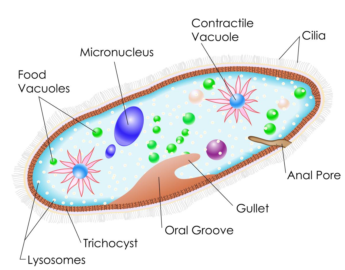
Examples of Protists - Biology Wise
Label the Microscope Diagram | Download Scientific Diagram the antibiogram of e. coli was investigated in different generations using eight antibiotic discs such as chloramphenicol (ch), streptomycin (s), gentamycin (g), ciprofloxacin (ci),...
Post a Comment for "43 simple microscope diagram with labels"