41 the human heart and its labels
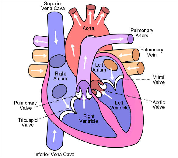
Human heart: Anatomy, function & facts | Live Science The human heart has four chambers: two upper chambers (the atria) and two lower ones (the ventricles), according to the National Institutes of Health. The right atrium and right ventricle together... Heart: Anatomy and Function - Cleveland Clinic A layer of muscular tissue called the septum divides your heart walls into the left and right sides. Your heart walls have three layers: Endocardium: Inner layer. Myocardium: Muscular middle layer. Epicardium: Protective outer layer. The epicardium is one layer of your pericardium. The pericardium is a protective sac that covers your entire heart.
Human Heart (Anatomy): Diagram, Function, Chambers, Location in ... - WebMD The heart is a muscular organ about the size of a fist, located just behind and slightly left of the breastbone. The heart pumps blood through the network of arteries and veins called the...

The human heart and its labels
Anatomy of a Human Heart - U of M Health The heart is made up of four chambers: two upper chambers known as the left atrium and right atrium and two lower chambers called the left and right ventricles. MORE FROM MICHIGAN: Sign up for our weekly newsletter It is also made up of four valves: the tricuspid, pulmonary, mitral and aortic valves. A Diagram of the Heart and Its Functioning Explained in Detail The heart blood flow diagram (flowchart) given below will help you to understand the pathway of blood through the heart.Initial five points denotes impure or deoxygenated blood and the last five points denotes pure or oxygenated blood. 1.Different Parts of the Body ↓ 2.Major Veins ↓ 3.Right Atrium ↓ 4.Right Ventricle ↓ 5.Pulmonary Artery ↓ 6.Lungs Human Heart - Diagram and Anatomy of the Heart - Innerbody The heart contains 4 chambers: the right atrium, left atrium, right ventricle, and left ventricle. The atria are smaller than the ventricles and have thinner, less muscular walls than the ventricles. The atria act as receiving chambers for blood, so they are connected to the veins that carry blood to the heart.
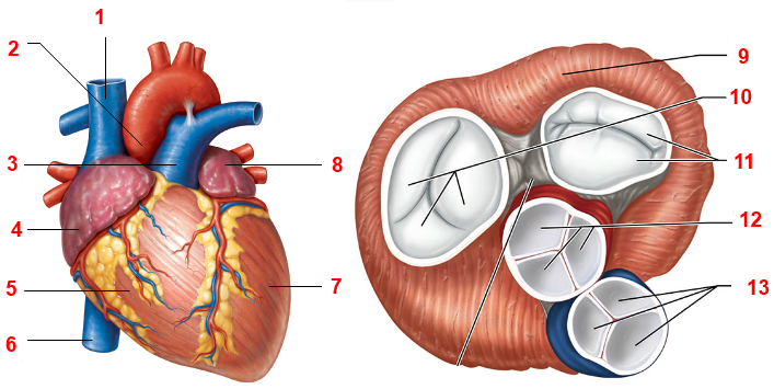
The human heart and its labels. Heart Diagram with Labels and Detailed Explanation - BYJUS Well-Labelled Diagram of Heart The heart is made up of four chambers: The upper two chambers of the heart are called auricles. The lower two chambers of the heart are called ventricles. The heart wall is made up of three layers: The outer layer of the heart wall is called epicardium. The middle layer of the heart wall is called myocardium. 147 Heart Anatomy With Labels Premium High Res Photos - Getty Images Browse 147 heart anatomy with labels stock photos and images available, or start a new search to explore more stock photos and images. of 3. NEXT. Human Circulatory System - Organs, Diagram and Its Functions - BYJUS The human heart consists of four chambers - two ventricles and two auricles. The human circulatory system possesses a body-wide network of blood vessels. These comprise arteries, veins, and capillaries. The primary function of blood vessels is to transport oxygenated blood and nutrients to all parts of the body. human system with label Human Male Skeleton - Stock Image - C024/9740 - Science Photo Library . Label The Human Stomach On A Diagram The Four Main Regions Of Stomach medicinebtg.com. stomach label anatomy diagram digestive human sphincter labeled regions system physiology identify curvatures its google tract body models types four
Human Heart - Anatomy, Functions and Facts about Heart - BYJUS The human heart is divided into four chambers, namely two ventricles and two atria. The ventricles are the chambers that pump blood and atrium are the chambers that receive the blood. Among which, the right atrium and ventricle make up the "right portion of the heart", and the left atrium and ventricle make up the "left portion of the heart." 5. The Anatomy of the Heart, Its Structures, and Functions - ThoughtCo The heart is made up of four chambers: Atria: Upper two chambers of the heart. Ventricles: Lower two chambers of the heart. Heart Wall The heart wall consists of three layers: Epicardium: The outer layer of the wall of the heart. Myocardium: The muscular middle layer of the wall of the heart. Endocardium: The inner layer of the heart. Label the heart — Science Learning Hub In this interactive, you can label parts of the human heart. Drag and drop the text labels onto the boxes next to the diagram. Selecting or hovering over a box will highlight each area in the diagram. pulmonary vein semilunar valve right ventricle right atrium vena cava left atrium pulmonary artery aorta left ventricle Download Exercise Tweet A Labeled Diagram of the Human Heart You Really Need to See The human heart, comprises four chambers: right atrium, left atrium, right ventricle and left ventricle. The two upper chambers are called the left and the right atria, and the two lower chambers are known as the left and the right ventricles. The two atria and ventricles are separated from each other by a muscle wall called 'septum'.
A Look at the Human Heart - ACLS The human heart is an incredible organ that plays a vital role in how your body works. It works like a pump which pushes life-giving blood through your body. The heart is the center of the circulatory system which feeds oxygenated blood, deoxygenated blood and nutrients to vital organs and muscle tissue throughout your body; it also helps to ... File:Diagram of the human heart (no labels).svg File:Diagram of the human heart (no labels).svg. From Wikimedia Commons, the free media repository. File. File history. File usage on Commons. Metadata. Size of this PNG preview of this SVG file: 498 × 599 pixels. Other resolutions: 199 × 240 pixels | 399 × 480 pixels | 639 × 768 pixels | 851 × 1,024 pixels | 1,703 × 2,048 pixels | 533 × ... Draw a sectional view of the human heart and label the aorta, right ... Draw a diagram to show the external structure of the human heart and label its various parts Mention the path by which the deoxygenated blood from the intestine travels up to the lungs. Hard. View solution > Draw the diagram showing the sectional view of the human heart. Label the following parts Heart: illustrated anatomy - e-Anatomy - IMAIOS This interactive atlas of human heart anatomy is based on medical illustrations and cadaver photography. The user can show or hide the anatomical labels which provide a useful tool to create illustrations perfectly adapted for teaching. Anatomy of the heart: anatomical illustrations and structures, 3D model and photographs of dissection.
Labeling the Heart (Part One) Quiz - By dilatory
Parts Of The Human Heart | Science Trends It is between the lungs, approximately in the middle of the chest, right behind the sternum (breastbone) but slightly to the left. The human heart beats (contracts) each time it received an electrical impulse from the heart muscle, known as the myocardium. The human heart together with the circulatory system make up the cardiovascular system.

Human Heart Poster | Zazzle.com | Human heart, Heart poster, Human body systems
13 parts of the human heart (and its functions) - LORECENTRAL Its opening (generated by the systole of the atrium) causes blood to travel between both regions. 3. Left Ventricle. Another major part of the heart. The left ventricle receives oxygen-rich blood from the left atrium and sends it to the rest of the body through the aortic artery. 4. Aortic sigmoid valve.

File:Diagram of the human heart (cropped).svg - Wikibooks, open books for an open world
Human Heart - Diagram and Anatomy of the Heart - Innerbody The heart contains 4 chambers: the right atrium, left atrium, right ventricle, and left ventricle. The atria are smaller than the ventricles and have thinner, less muscular walls than the ventricles. The atria act as receiving chambers for blood, so they are connected to the veins that carry blood to the heart.

Biology Diagrams,Images,Pictures of Human anatomy and physiology : Lymphatic System
A Diagram of the Heart and Its Functioning Explained in Detail The heart blood flow diagram (flowchart) given below will help you to understand the pathway of blood through the heart.Initial five points denotes impure or deoxygenated blood and the last five points denotes pure or oxygenated blood. 1.Different Parts of the Body ↓ 2.Major Veins ↓ 3.Right Atrium ↓ 4.Right Ventricle ↓ 5.Pulmonary Artery ↓ 6.Lungs

Pin on Classical Conversations Science
Anatomy of a Human Heart - U of M Health The heart is made up of four chambers: two upper chambers known as the left atrium and right atrium and two lower chambers called the left and right ventricles. MORE FROM MICHIGAN: Sign up for our weekly newsletter It is also made up of four valves: the tricuspid, pulmonary, mitral and aortic valves.


give it some heart on Pinterest | Human Heart, Anatomical Heart and Anatomy
human heart without label - Clip Art Library
ANATOMY OF THE HUMAN HEART Medical Science Educational Wall Chart 24x36 POSTER | eBay
Heart Diagram – 15+ Free Printable Word, Excel, EPS, PSD Template Download | Free & Premium ...


35 Label This Anterior View Of The Human Heart - Labels Design Ideas 2020

NCERT Class 10 Chapter 7 Life Process CBSE Board Sample Problems Long Answer- FlexiPrep

Dazzling Wings: 2013

Abstract Art Pictures Collection: Human Eyes Captured!

23 Heart-Melting Pictures Of Animals Who Love Each Other Too Much

35 Heart Anatomy Quiz Label - Labels Information List
Post a Comment for "41 the human heart and its labels"